

ISSN 1004-5759 CN 62-1105/S
草业学报 ›› 2021, Vol. 30 ›› Issue (10): 92-104.DOI: 10.11686/cyxb2020360
杨凯( ), 史娟(), 袁玉涛, 王立婷
收稿日期:2020-07-28
修回日期:2020-09-27
出版日期:2021-09-16
发布日期:2021-09-16
通讯作者:
史娟
作者简介:Corresponding author. E-mail: shi_j@nxu.edu.cn基金资助:
Kai YANG(), Juan SHI(), Yu-tao YUAN, Li-ting WANG
Received:2020-07-28
Revised:2020-09-27
Online:2021-09-16
Published:2021-09-16
Contact:
Juan SHI
摘要:
为了揭示白三叶草和白粉菌互作的细胞学机制,明确白三叶草白粉菌的分类地位,为牧草抗病育种和科学防控提供依据。采用光学显微镜和透射电镜技术观察了白粉菌侵入白三叶草叶片感染白粉病的细胞生理特征。通过传统形态学和ITS序列分析对白三叶草白粉病病原菌进行了鉴定,采用水琼脂玻片法观察了病原菌分生孢子萌发特性。结果表明,白粉菌在白三叶草表皮细胞和保卫细胞交界处直接侵入,菌丝侵入表皮细胞壁与寄主接触部分的胞壁组织被降解且颜色加深,对应的细胞内侧产生大量胞壁沉积物,细胞壁不再完整气孔通道变形,保卫细胞发生质膜分离;进入表皮细胞的吸器被寄主质膜包围形成交界面,栅栏组织细胞内叶绿体肿胀,由椭圆形变为近球形聚集,淀粉粒由长条形变为椭圆形,嗜锇颗粒增多。接种到叶片的分生孢子4 h萌发长出初生芽管,10 h形成附着胞,12 h附着胞一侧形成侵染钉,48 h次生菌丝形成,96 h时形成大量菌丝并分化出成熟的分生孢子梗,叶片显症;144 h时分生孢子梗分化出2~3个分生孢子,顶端最先成熟,168 h时粉层扩展至叶片的2/3,240 h时达到90%以上,且粉层加厚,叶片卷曲。依据分生孢子的形态,结合ITS序列鉴定,将宁夏地区白三叶草白粉病的病原菌鉴定为豌豆白粉菌。该菌在25 ℃、 pH为7.0时生长最好,光照条件有利于分生孢子萌发。
杨凯, 史娟, 袁玉涛, 王立婷. 白三叶草叶片感染白粉病的细胞生理变化及其病原鉴定[J]. 草业学报, 2021, 30(10): 92-104.
Kai YANG, Juan SHI, Yu-tao YUAN, Li-ting WANG. Pathogen identification and cell physiological changes of Trifolium repens leaves infected with powdery mildew[J]. Acta Prataculturae Sinica, 2021, 30(10): 92-104.

图1 白粉病菌危害白三叶草叶片症状A: 健康的白三叶草; B: 接种白粉菌4 d的叶片表面出现稀疏白粉层; C: 接种5 d,白粉层加厚; D~E: 接种6~7 d,粉斑扩展至叶片的1/3~2/3; F: 接种10 d,粉层覆盖率达到90%以上,叶片卷曲; G~H: 接种15~20 d,整个叶片粉层加厚,叶片变黄,萎蔫,并逐渐枯死。 A: Healthy T. repens; B: The leaves inoculated with powdery mildew for 4 d had sparse powdery layers on the surface; C: The white powder layer was thickened after inoculation for 5 d; D-E: After inoculation for 6-7 d, the powder spots extended to 1/3-2/3 of the leaves; F: The coverage rate of powder layer reached more than 90% after inoculation for 10 d, and the leaves were curled; G-H: When inoculated to 15-20 d, the entire leaf powder layer thickened, the leaves became yellow, shriveled, and gradually withered.
Fig. 1 Symptoms of leaf damage of T. repens by E. pisi
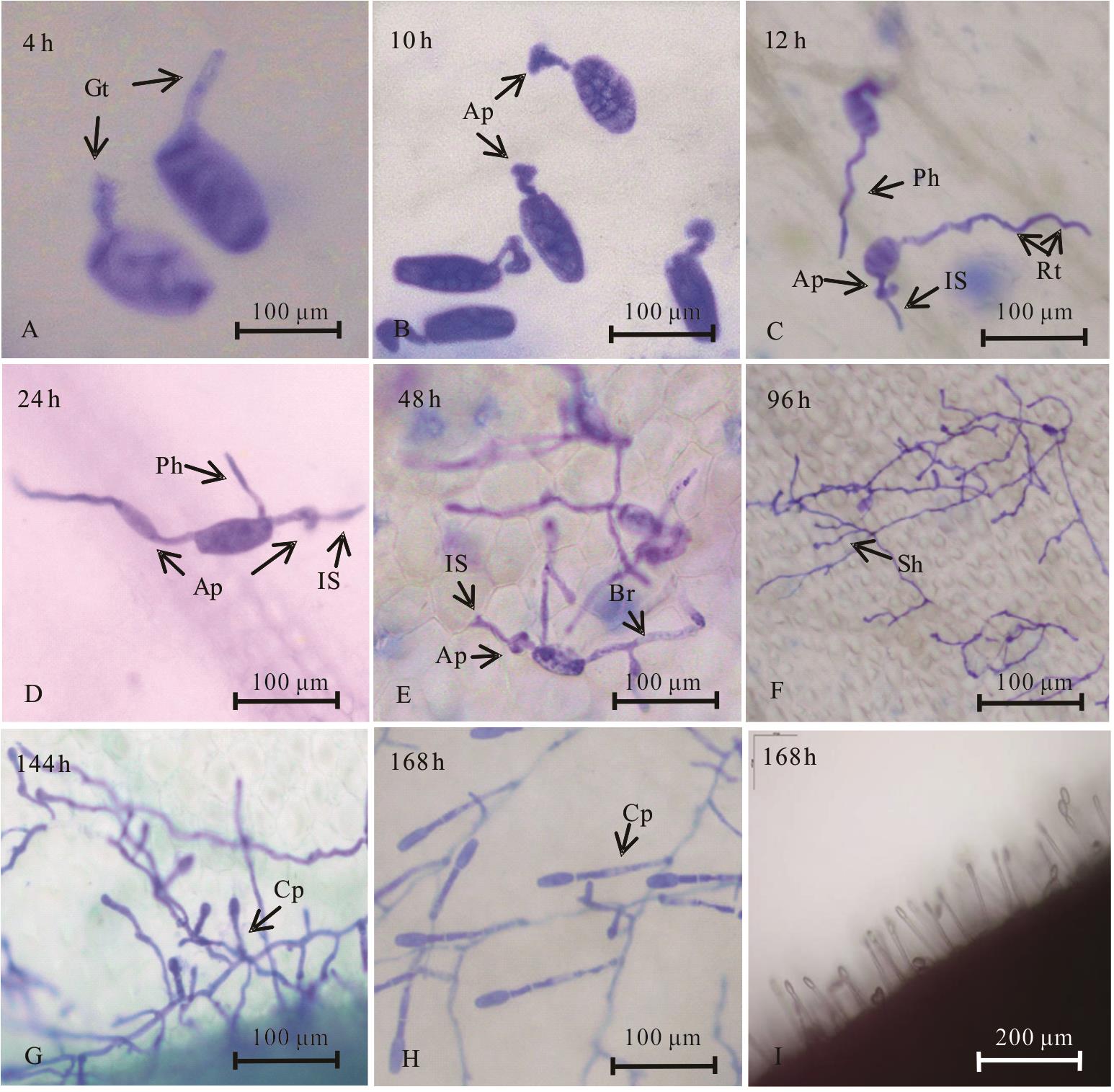
图2 白粉病菌在白三叶草叶片的发育情况A: 4 h时分生孢子顶端肩胛处长出芽管;B:10 h时附着胞形成; C: 12 h时侵染钉形成,且芽管生长形成拱状菌丝,拱状菌丝基部为产生吸器部位;D: 24 h时分生孢子变扁且芽管基部长出初生菌丝;E:48 h时次生菌丝形成; F: 96 h时菌丝形成网状; G: 144 h时孢子梗形成; H: 168 h时每个孢子梗串生2 ~ 3个分生孢子;I:在解剖镜下将叶片对折侧面观察到的分生孢子梗; Gt: 芽管; Ap: 附着胞; IS: 侵染钉; Ph: 初生菌丝; Br: 分枝 ;Sh: 次生菌丝; Cp: 叶片表面的分生孢子梗; Rt: 拱形菌丝基部。 A: At 4 h, the germinal tube appears at the apex of the spore scapula; B: Appressorium formation at 10 h; C: At 12 h, the infection nail was formed, and the bud tube grew to form an arch hypha. The base of the arch hypha was the site of producing haustorium; D: At 24 h, the germinal spore flattens and the primary hypha grows out of the bud; E: Secondary hyphae formed at 48 h; F: Hyphae formed a network at 96 h; G: Sporoderm formation at 144 h; H: At 168 h, 2-3 conidia were born on each spore stem; I: Conidiospore peduncles observed on the folded side of the leaf under anatomic microscope; Gt: Germ tube; Ap: Appressorium; IS: Invasion site; Ph: Primary hyphae; Br: Branch; Sh: Secondary hyphae; Cp: Conidia peduncle; Rt: Root.
Fig. 2 Development of E. pisi in T. repens

图3 正常白三叶草寄主组织与受白粉病菌侵入寄主组织的半薄切片A: 正常白三叶草表皮组织;B:受白粉菌侵染的白三叶草表皮组织; Ue: 上表皮; De: 下表皮; Pt: 栅栏组织; St: 海绵组织; Vb: 维管束; Hs: 吸器; Hp: 菌丝。 A: Normal T. repens epidermal tissue; B: Epidermal tissue of T. repens infected by powdery mildew; Ue: Upper epidermis; De:Down epidermis; Pt: Palisade tissue; St: Sponge tissue; Vb: Vascular bundle; Hs: Haustoria; Hp: Hyphae.
Fig. 3 A semi-thin section of normal T. repens host tissue and powdery mildew invaded the host tissue

图4 病原菌侵入寄主细胞的超微结构特征A: 菌丝内大量的细胞器及一侧形成凸起;B:菌丝内的线粒体及核糖体; C: 菌丝凸起且具有膜性物; D: 有隔菌丝;E:有隔菌丝内的细胞器; F: 白粉菌菌丝侵入寄主前,寄主响应侵染结构的变化; G: 白粉菌侵入寄主时,菌丝与寄主发生的系列变化; H~I: 侵入菌丝对应的表皮细胞内发生的变化; J: 表皮细胞内形成了吸器;K:吸器内细胞器及外壁被一层厚膜包围; L~M: 受白粉菌侵染的白三叶草表皮细胞内叶绿体结构发生变化; N:正常的叶绿体结构; O: 正常的白三叶草表皮细胞; Nu: 细胞核; Mi: 线粒体; Bv: 大液泡; Ri: 核糖体; Ch: 叶绿体; Hs: 吸器;V e: 囊泡; S: 淀粉粒; P: 嗜锇颗粒。 A: A large number of organelles and one side of hyphae form A bulge; B: Mitochondria and ribosomes in hyphae; C: Hypha is convex and has membranous properties; D: There are septal hyphae; E: There are organelles in the septal hyphae; F: The host responds to the change of the infective structure before the invasion of mycelia; G: Series changes of mycelia and host when powdery mildew invades the host; H-I: Changes in epidermal cells corresponding to invasion of mycelia; J: Haustors form inside the epidermal cells; K: The inner organelle and outer wall of the haustoria are surrounded by a thick film; L-M: Chloroplast structure changes in epidermal cells of the infected white clover; N: Normal chloroplast structure; O: Normal white clover epidermal cells; Nu: Nucleus; Mi: Mitochondria; Bv: Big vacuole; Ri: Ribosome; Ch: Chloroplast; Hs: Haustoria; Ve: Vesica; S: Starch granule; P: Plastoglobule.
Fig. 4 Ultrastructural characteristics of pathogen invading host cells
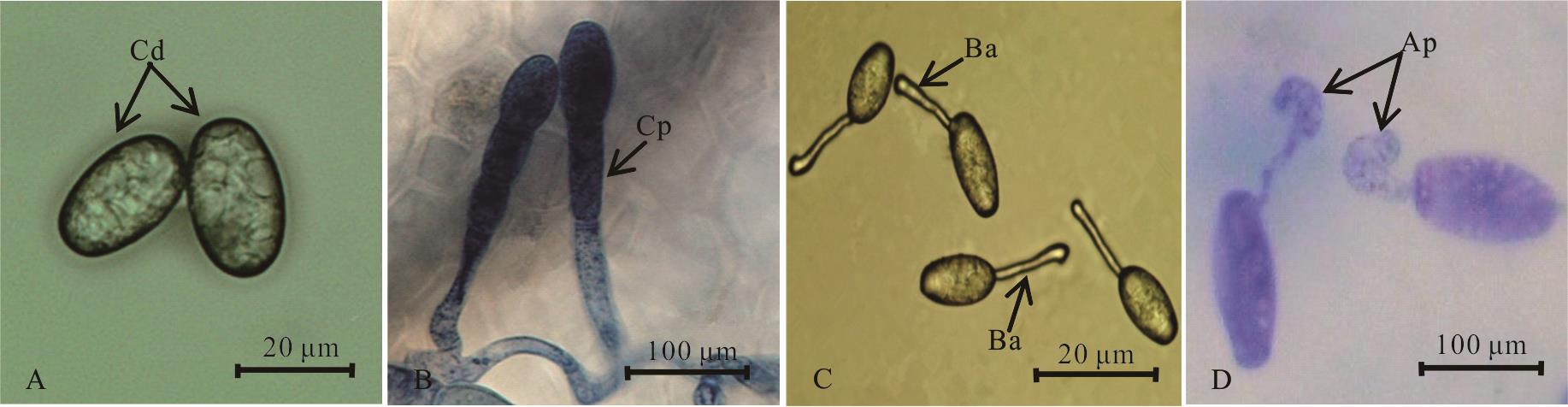
图5 白三叶白粉病病原菌的形态特征A: 白粉菌分生孢子;B:分生孢子梗; C: 水琼脂玻片下观察到的分生孢子顶部芽管生长; D: 叶片上观察到分生孢子萌发产生芽管并形成附着胞;Cd:分生孢子; Cp: 分生孢子梗; Ba: 芽管及顶端膨大; Ap: 附着胞。 A: Conidia of powdery mildew; B: Conidia peduncle; C: Conidia sprout tube observed under water agar slide; D: Conidia germination was observed on the leaves to produce bud tubes and appressorium; Cd: Conidia; CP: Conidiophore; Ba: Bud tube and apex inflated; Ap: Appressorium.
Fig.5 Morphological characteristics of fungus causing powdery mildews on T. repens
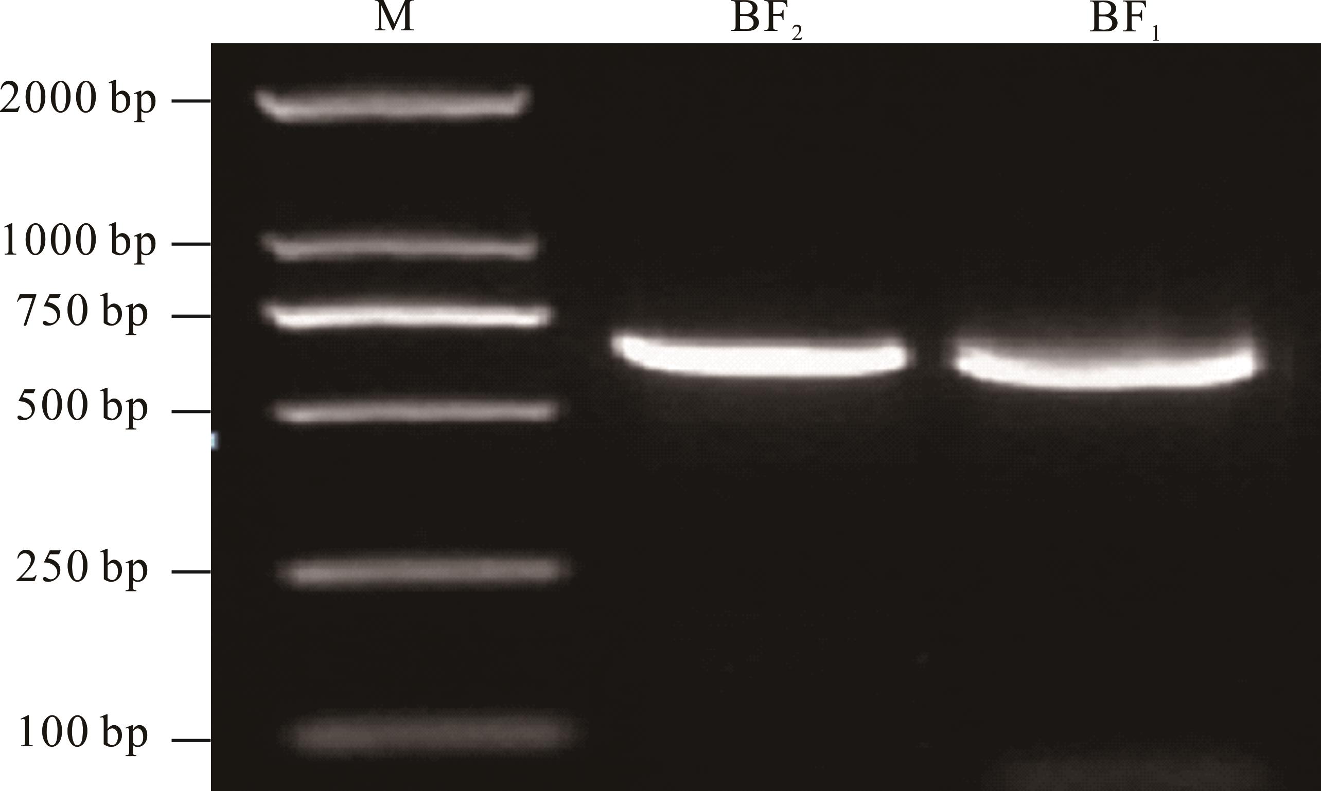
图6 白粉病菌ITS序列片段的PCR扩增
Fig.6 PCR amplification of ribosomal ITS sequence segments of powdery mildew

图7 ITS序列构建的系统发育树
Fig.7 Phylogenetic tree constructed by ITS sequence
温度 Temperature (℃) | 孢子萌发率 Spore germination rate (%) | ||||
|---|---|---|---|---|---|
| 4 h | 6 h | 8 h | 12 h | 24 h | |
| 5 | 0.53C | 2.17C | 2.97B | 5.45B | 7.55B |
| 10 | 0.98C | 4.77C | 8.27B | 16.00B | 17.43B |
| 15 | 7.01B | 19.00B | 27.40A | 33.15A | 40.47A |
| 20 | 9.42AB | 18.55B | 21.50A | 32.29A | 35.85A |
| 25 | 11.57A | 28.21A | 31.25A | 37.76A | 47.53A |
| 28 | 2.54C | 8.80C | 9.63B | 14.83B | 18.65B |
| 30 | 0.97C | 4.62C | 6.78B | 7.19B | 9.15B |
表1 不同温度对白三叶草白粉菌分生孢子萌发的影响
Table 1 Effects of different temperatures on the conidia germination of powdery mildew in T. repens
温度 Temperature (℃) | 孢子萌发率 Spore germination rate (%) | ||||
|---|---|---|---|---|---|
| 4 h | 6 h | 8 h | 12 h | 24 h | |
| 5 | 0.53C | 2.17C | 2.97B | 5.45B | 7.55B |
| 10 | 0.98C | 4.77C | 8.27B | 16.00B | 17.43B |
| 15 | 7.01B | 19.00B | 27.40A | 33.15A | 40.47A |
| 20 | 9.42AB | 18.55B | 21.50A | 32.29A | 35.85A |
| 25 | 11.57A | 28.21A | 31.25A | 37.76A | 47.53A |
| 28 | 2.54C | 8.80C | 9.63B | 14.83B | 18.65B |
| 30 | 0.97C | 4.62C | 6.78B | 7.19B | 9.15B |
光照时间 Illumination treatment | 孢子萌发率Spore germination rate | ||||
|---|---|---|---|---|---|
| 4 h | 6 h | 8 h | 12 h | 24 h | |
| 全光照 Full illumination | 20.06A | 36.88A | 40.75A | 43.40A | 45.82A |
| 光暗交替 Half illumination | 13.90AB | 27.54A | 30.12A | 36.66A | 39.96A |
| 全黑暗 Total darkness | 8.33B | 20.98A | 27.03A | 30.17A | 33.87A |
表2 不同光照对白三叶草白粉菌分生孢子萌发的影响
Table 2 Effect of different lights on the conidia germination of powdery mildew in T. repens (%)
光照时间 Illumination treatment | 孢子萌发率Spore germination rate | ||||
|---|---|---|---|---|---|
| 4 h | 6 h | 8 h | 12 h | 24 h | |
| 全光照 Full illumination | 20.06A | 36.88A | 40.75A | 43.40A | 45.82A |
| 光暗交替 Half illumination | 13.90AB | 27.54A | 30.12A | 36.66A | 39.96A |
| 全黑暗 Total darkness | 8.33B | 20.98A | 27.03A | 30.17A | 33.87A |
| pH | 孢子萌发率Spore germination rate | ||||
|---|---|---|---|---|---|
| 4 h | 6 h | 8 h | 12 h | 24 h | |
| 4.0 | 2.75CD | 10.87BC | 12.54C | 15.20B | 15.78B |
| 5.0 | 2.94CD | 11.12BC | 14.25B | 19.66B | 25.82B |
| 6.0 | 11.08B | 17.73AB | 23.20B | 32.43A | 37.51A |
| 7.0 | 16.86A | 24.77A | 35.97A | 40.99A | 45.50A |
| 8.0 | 8.01BC | 15.02B | 23.11B | 31.56A | 37.12A |
| 9.0 | 2.13D | 4.95C | 7.50C | 12.75B | 18.72B |
表3 不同pH对白三叶草白粉菌分生孢子萌发的影响
Table 3 Effects of different pH on the conidia germination of powdery mildew in T. repens (%)
| pH | 孢子萌发率Spore germination rate | ||||
|---|---|---|---|---|---|
| 4 h | 6 h | 8 h | 12 h | 24 h | |
| 4.0 | 2.75CD | 10.87BC | 12.54C | 15.20B | 15.78B |
| 5.0 | 2.94CD | 11.12BC | 14.25B | 19.66B | 25.82B |
| 6.0 | 11.08B | 17.73AB | 23.20B | 32.43A | 37.51A |
| 7.0 | 16.86A | 24.77A | 35.97A | 40.99A | 45.50A |
| 8.0 | 8.01BC | 15.02B | 23.11B | 31.56A | 37.12A |
| 9.0 | 2.13D | 4.95C | 7.50C | 12.75B | 18.72B |
| 1 | Wang Z Y, Shu J H, Chen Y, et al.Analysis of Trifolium repens DNA methylation of Guizhou different regions by MSAP. Genomics and Applied Biology, 2020, 39(4): 1741-1750. |
| 王子苑, 舒健虹, 陈莹, 等. 贵州不同地区白三叶基因组甲基化MSAP分析. 基因组学与应用生物学, 2020, 39(4): 1741-1750. | |
| 2 | Huang Y M, Zhang K, Sun L X, et al.Effects of Trifolium repens invasion on soil animals in an urban turf ecosystem. Acta Ecologica Sinica, 2018, 38(23): 8489-8499. |
| 黄玉梅, 张凯, 孙凌霞, 等. 白三叶(Trifolium repens)入侵对城市草坪生态系统土壤动物的影响. 生态学报, 2018, 38(23): 8489-8499. | |
| 3 | Liang Z, Jiang S J, Wei L, et al. Progress in research on Trifolium genetic engineering. Acta Prataculturae Sinica, 2009, 18(2): 205-211. |
| 梁哲, 姜三杰, 未丽, 等. 三叶草基因工程研究进展. 草业学报, 2009, 18(2): 205-211. | |
| 4 | Chen B S. Forage crop cultivation. Beijing: China Agriculture Press, 2001: 224-233. |
| 陈宝书. 牧草饲料作物栽培学. 北京: 中国农业出版社, 2001: 224-233. | |
| 5 | Li Y X, Zhang T B, Du H L, et al. The allelopathy of five pine needle extracts on Trifolium repens L. Journal of Nuclear Agricultural Sciences, 2020, 34(7): 1606-1612. |
| 李雅馨, 张天宝, 杜慧玲, 等. 5种松针浸提液对白三叶草的化感作用. 核农学报, 2020, 34(7): 1606-1612. | |
| 6 | Jin Z M, Sha W. Study on drought resistance of Trifolium repens Linn seedlings. Northern Horticulture, 2010(18): 50-52. |
| 金忠民, 沙伟. 白三叶抗旱生理的研究. 北方园艺, 2010(18): 50-52. | |
| 7 | Li Z, Peng Y, Yin S X, et al. Effects of exogenous mannose application on drought tolerance, sugars, and sugar alcohol accumulation in white clover. Acta Prataculturae Sinica, 2019, 28(12): 85-93. |
| 李州, 彭燕, 尹淑霞, 等. 甘露糖对白三叶抗旱性、糖及糖醇类代谢物积累的影响. 草业学报, 2019, 28(12): 85-93. | |
| 8 | Qu L L, Wang J J. Research progress on stress resistance of Trifolium repens. Chinese Journal of Grassland, 2020, 42(2): 155-161. |
| 屈璐璐, 王俊杰. 白三叶抗逆性研究进展. 中国草地学报, 2020, 42(2): 155-161. | |
| 9 | Lan H Y. Common diseases and insect pests of herbage and their control methods. Contemporary Animal Husbandry, 2018(26): 17-18. |
| 兰惠英. 牧草常见病虫害及防治方法. 当代畜牧, 2018(26): 17-18. | |
| 10 | Liu X L, Song C, Du W H. Study on mechanism of resistance to powdery mildew in Minshan red clover. Grassland and Turf, 2011, 31(3): 73-76. |
| 刘晓玲, 宋超, 杜文华. 红三叶对白粉病抗性机理研究. 草原与草坪, 2011, 31(3): 73-76. | |
| 11 | Yang F. Identification and prevention of powdery mildew in garden lawn. Xinjiang Agricultural Science and Technology, 2008(3): 62. |
| 杨芬. 园林草坪白粉病的识别与防治. 新疆农业科技, 2008(3): 62. | |
| 12 | Sang W J, Liang J, Li Y S. Investigation on powdery mildew inlidence of Trifolium pratense in Guiyang. Journal of Southwest University (Natural Science Edition), 1990(4): 353-355. |
| 桑维均, 梁建, 李永顺. 贵阳地区红三叶草白粉病病情消长和病菌生物学特性. 西南农业大学学报(自然科学版), 1990(4): 353-355. | |
| 13 | Yuan Y T, Shi J, Ma X, et al. Identification and biological characteristics of alfalfa powdery mildew fungi. Microbiology China, 2020, 47(11): 3539-3550. |
| 袁玉涛, 史娟, 马新, 等. 紫花苜蓿白粉病病原菌鉴定及其生物学特性. 微生物学通报, 2020, 47(11): 3539-3550. | |
| 14 | Li S, Zhu L L, Li J F, et al. Identification of the pathogen causing tomato powdery mildew in Heilongjiang Province. Plant Protection, 2014, 40(4): 112-114, 129. |
| 李帅, 朱路路, 李景富, 等. 黑龙江省番茄白粉病病原鉴定. 植物保护, 2014, 40(4): 112-114, 129. | |
| 15 | Zheng L, Wu X Q.Advances on infection structures of plant pathogenic fungi. Journal of Nanjing Forestry University (Natural Sciences Edition) , 2007(1): 90-94. |
| 郑玲, 吴小芹. 植物病原真菌侵染结构研究进展. 南京林业大学学报(自然科学版), 2007(1): 90-94. | |
| 16 | Riopel J L. Haustorial initiation and differentiation. New York: Parasitic Plants, Chapman and HallPress, 1995. |
| 17 | Ren B, Gao X N, Han Q M, et al. Etiology and infection process of Glomerella cingulata causing Glomerella leaf spot of apple. Acta Phytophylacica Sinica, 2014, 41(5): 608-614. |
| 任斌, 高小宁, 韩青梅, 等. 苹果炭疽叶枯病病原(Glomerella cingulata)及其侵染过程. 植物保护学报, 2014, 41(5): 608-614. | |
| 18 | Yang R L, Liu J Y, Lv X, et al.Micro-and ultrastructural changes of wheat leaf cells induced by powdery mildew infection. Acta Botanica Boreali-Occidentalia Sinica, 2001(2): 293-296, 400-401. |
| 杨若林, 刘建云, 吕欣, 等. 白粉菌侵染对小麦叶片显微及超微结构的影响. 西北植物学报, 2001(2): 293-296, 400-401. | |
| 19 | Zhang Z L, Qu W J. Experimental instruction in plant physiology (3rd Edition). Beijing: Higher Education Press, 2003: 206. |
| 张志良, 瞿伟菁. 植物生理学实验指导(第3版). 北京: 高等教育出版社, 2003: 206. | |
| 20 | Kang Z S. Ultrastructure of plant pathogenic fungi. Beijing: China Science and Technology Press, 1996: 1-74. |
| 康振生. 植物病原真菌超微结构. 北京: 中国科学技术出版社, 1996: 1-74. | |
| 21 | Li W X, Xiao R G, Lv M M, et al. Establishment and application of real-time PCR for quantitatively detecting Plasmopara viticola in Vitis vinifera. Scientia Agricultura Sinica, 2019, 52(9): 1529-1540. |
| 李文学, 肖瑞刚, 吕苗苗, 等. 葡萄霜霉病菌实时荧光定量PCR检测体系的建立和应用. 中国农业科学, 2019, 52(9): 1529-1540. | |
| 22 | Fang Z D. Research methods of plant diseases. Beijing: China Agriculturae Press, 1998: 140-145. |
| 方中达. 植病研究方法. 北京: 中国农业出版社, 1998: 140-145. | |
| 23 | Wei J C. Fungus identification manual. Shanghai: Shanghai Scientific & Technical Publishers, 1979: 1-9. |
| 魏景超. 真菌鉴定手册. 上海: 上海科学技术出版社, 1979: 1-9. | |
| 24 | Zheng R Y, Yu Y N. Flora of fungi in China, Volume I, powdery mildew. Beijing: Science Press, 1987: 5-13. |
| 郑儒永, 余永年. 中国真菌志, 第一卷, 白粉菌目. 北京: 科学出版社, 1987: 5-13. | |
| 25 | Bushnell W R. Aggregation of host cytoplasm and the formation of papillae and haustoria in powdery mildew of barley. Phytopathology, 1975, 65: 310-318. |
| 26 | Wan S L, Liang P, Liu W B, et al.Cytological analysis of compatible interactions between rubber tree and Oidium heveae. Plant Protection, 2014, 40(3): 26-36. |
| 万三连, 梁鹏, 刘文波, 等. 橡胶树与白粉病菌(Oidium heveae)亲和互作组织细胞学研究. 植物保护, 2014, 40(3): 26-36. | |
| 27 | Zhang Y M, Ma H L, Tang Y Z. Structural changes in leaves in Medicago sativa infected with Erysiphe pisi. Acta Prataculturae Sinica, 2017, 26(2): 88-94. |
| 张咏梅, 马晖玲, 唐云智. 紫花苜蓿叶片受白粉病菌侵染后结构的变化. 草业学报, 2017, 26(2): 88-94. | |
| 28 | Lu Q, Yang L, Wang H W, et al.Responses of photosynthetic characteristics and chloroplast ultrastructure to salt stress in seedlings of Cornus hongkongensis subsp. elegans. Journal of Nanjing Forestry University (Natural Sciences Edition), 2020, 44(4): 29-36. |
| 鲁强, 杨玲, 王昊伟, 等. 秀丽四照花光合特性和叶绿体超微结构的盐胁迫响应. 南京林业大学学报(自然科学版), 2020, 44(4): 29-36. | |
| 29 | Li X F, Ni Z M, Wu Y Y, et al.Effects of salt stress on photosynthetic characteristics and leaf cell structure of ‘Yinhong’ grape seedlings. Acta Ecologica Sinica, 2015, 35(13): 4436-4444. |
| 李学孚, 倪智敏, 吴月燕, 等. 盐胁迫对‘鄞红’葡萄光合特性及叶片细胞结构的影响. 生态学报, 2015, 35(13): 4436-4444. | |
| 30 | Kirchhoff H . Chloroplast ultrastructure in plants. New Phytologist, 2019, 223(2): 565-574. |
| 31 | Van Wijk K J, Kessler F. Plastoglobuli: Plastid microcompartments with integrated functions in metabolism, plastid developmental transitions, and environmental adaptation. Annual Review of Plant Biology, 2017, 68(1): 253-289. |
| 32 | Liu M, Fang Y L. Effects of heat stress on physiological indexes and ultrastructure of grapevines. Scientia Agricultura Sinica, 2020, 53(7): 1444-1458. |
| 刘敏, 房玉林. 高温胁迫对葡萄幼树生理指标和超显微结构的影响. 中国农业科学, 2020, 53(7): 1444-1458. | |
| 33 | Xu W, Wang Y, Yuan Q H, et al. Effect of NaCl on ultrastructure in leaves of white clovers (Trifolium repens L.). Chinese Journal of Grassland, 2013, 35(6): 104-108. |
| 徐威, 王瑜, 袁庆华, 等. NaCl胁迫对白三叶叶片超微结构的影响. 中国草地学报, 2013, 35(6): 104-108. | |
| 34 | Shi J, Wang H R, Zhong S L.Ultrastructural characteristics of compatible Pseudopeziza medicaginis interaction with alfalfa leaf. Acta Prataculturae Sinica, 2012, 21(5): 122-127. |
| 史娟, 王华荣, 钟少林. 苜蓿假盘菌与苜蓿叶片亲和性互作的超微结构特征. 草业学报, 2012, 21(5): 122-127. | |
| 35 | Chen S H, Lu Z Q. Occurrence and control measures of powdery mildew of red clover in artificial pasture. Journal of Grassland and Forage Science, 1997(2): 47-48. |
| 陈曙晖, 陆忠权. 人工草场红三叶草白粉病的发生与防治措施. 四川草原, 1997(2): 47-48. | |
| 36 | Yan Y, Li W P, Gao W J, et al. Application of rDNA ITS sequence analysis in fungus identification. Chinese Journal of Health Laboratory Technology, 2008(10): 1958-1961. |
| 燕勇, 李卫平, 高雯洁, 等. rDNA-ITS序列分析在真菌鉴定中的应用. 中国卫生检验杂志, 2008(10): 1958-1961. | |
| 37 | Wang L J, Guo N, Wang W, et al. The soil water content monitoring based on the temperature difference in the Northwest China. Research of Soil and Water Conservation, 2018, 25(5): 330-336. |
| 王丽娟, 郭铌, 王玮, 等. 基于温度差监测西北地区的土壤相对湿度. 水土保持研究, 2018, 25(5): 330-336. |
| [1] | 王春明, 元维伟, 张小杰, 周天旺, 郭成, 金社林. 二月兰叶斑病病原甘蓝链格孢的分离鉴定及生物学特性研究[J]. 草业学报, 2020, 29(5): 88-97. |
| [2] | 聂秀美, 赵桂琴, 孙浩洋, 柴继宽, 兰晓君, 周恒, 黎蓉, 琚泽亮, 焦润安, 孙雷雷. 甘肃省燕麦主产区叶斑病调查及病原鉴定[J]. 草业学报, 2020, 29(4): 157-167. |
| [3] | 程守丰, 梁巧兰, 魏列新, 桑旭文, 姜玉玲. 苜蓿不同品种AMV和WCMV带毒检测及生理生化特性研究[J]. 草业学报, 2020, 29(12): 140-149. |
| [4] | 陈斌, 李洪瑶, 刘筱玮, 夏斌, 孙绍文, 孙颖, 何淼. 不同光照强度对新娘草叶片形态建成及超微结构的影响[J]. 草业学报, 2019, 28(7): 175-185. |
| [5] | 孙海荣, 车昭碧, 陈乙实, 鲁为华, 王树林, 李娜娜, 辛怀璐. 荒漠植物囊果草生物学特性及其种群分布格局的生态适应意义[J]. 草业学报, 2019, 28(7): 198-207. |
| [6] | 李建宏, 李雪萍, 李昌宁, 韩冰, 徐万里, 姚拓. 一株植物根际促生菌Gnyt1的特性研究及分类地位的确定[J]. 草业学报, 2019, 28(5): 55-67. |
| [7] | 丁爱强, 徐先英, 张雯, 刘江, 富丽, 付贵全. 不同退化程度柽柳灌丛的土壤理化和生物学特性[J]. 草业学报, 2019, 28(2): 1-11. |
| [8] | 冯鹏, 孙力, 申晓慧, 李如来, 李增杰, 李志民, 郑海燕, 姜成, 杨鹤, 刘俊刚, 郭伟, 张英俊. 不同诱变处理对苜蓿叶片细胞显微和超微结构的影响[J]. 草业学报, 2018, 27(6): 72-80. |
| [9] | 蒲小剑,田久胜,田新会,杜文华. 红三叶遗传图谱构建及抗白粉病基因QTL定位[J]. 草业学报, 2018, 27(4): 79-88. |
| [10] | 董姬妃, 张帆, 胡雨寒, 李俊承, 李维, 王娴淑. 镉胁迫下增施氮对白三叶草生长的影响和镉毒害的缓解效应研究[J]. 草业学报, 2017, 26(9): 83-91. |
| [11] | 杨成德, 卞静, 陈泰祥, 陈秀蓉, 王涵琦, 杨小利, 王艳. 当归炭疽病菌的生物学特性研究[J]. 草业学报, 2017, 26(6): 139-144. |
| [12] | 贺春贵, 何振富, 王斐. 光敏型高丹草复种穴播高效栽培模式研究[J]. 草业学报, 2017, 26(5): 70-80. |
| [13] | 李健, 李美, 高兴祥, 房锋, 董连红. 菟丝子生防菌“鲁保一号”生物学特性及T-DNA插入突变体库的构建[J]. 草业学报, 2017, 26(1): 142-148. |
| [14] | 李健, 李美, 高兴祥, 房锋, 董连红. 稗草生防菌BC-1的分离及生物学特性研究[J]. 草业学报, 2016, 25(8): 164-171. |
| [15] | 赵富强, 张海琴, 孙宗华, 焦振飞, 刘晓燕, 陈韦寰, 陈国跃, 周永红. 鹅观草不同居群条锈病和白粉病抗性评价[J]. 草业学报, 2016, 25(4): 149-158. |
| 阅读次数 | ||||||
|
全文 |
|
|||||
|
摘要 |
|
|||||