ISSN 1004-5759 CN 62-1105/S

Acta Prataculturae Sinica ›› 2024, Vol. 33 ›› Issue (12): 188-200.DOI: 10.11686/cyxb2024024
Yue CHEN( ), Pin SONG, Man-man HOU, Xiao-ran YANG, Li-ping LIU, Ying-dong NI(
 )
)
Received:2024-01-16
Revised:2024-03-11
Online:2024-12-20
Published:2024-10-09
Contact:
Ying-dong NI
Yue CHEN, Pin SONG, Man-man HOU, Xiao-ran YANG, Li-ping LIU, Ying-dong NI. Effects of dietary supplementation with bile acid on ileal epithelial morphology, microflora composition, and relative transcript levels of IFN-γ in the ileal mucosa of goats with subacute ruminal acidosis[J]. Acta Prataculturae Sinica, 2024, 33(12): 188-200.
| 日粮组成Diet ingredient | 对照组CON (%) | 高精料组SARA (%) | 营养水平Nutrient level2) | 对照组CON | 高精料组SARA |
|---|---|---|---|---|---|
| 苜蓿草 Alfalfa | 67.45 | 28.91 | 代谢能 ME (MJ·kg-1) | 1.87 | 1.90 |
| 玉米 Corn | 20.41 | 57.03 | 粗蛋白 Crude protein (%) | 16.79 | 16.73 |
| 豆粕 Soybean meal | 8.50 | 10.42 | 中性洗涤纤维NDF (%) | 33.71 | 28.79 |
| 磷酸氢钙 Calcium hydrophosphate | 1.10 | 1.10 | 酸性洗涤纤维 ADF (%) | 18.22 | 14.32 |
| 石粉 Limestone | 1.20 | 1.20 | 粗灰分Ash (%) | 4.74 | 4.83 |
| 食盐 Salt | 0.40 | 0.40 | 粗脂肪 Ether extract (%) | 3.76 | 3.61 |
| 预混料 Premix1) | 0.94 | 0.94 | 淀粉Starch (%) | 24.25 | 31.21 |
| 总计 Total | 100.00 | 100.00 | 钙Calcium (%) | 0.95 | 0.96 |
| 磷Phosphorus (%) | 0.42 | 0.44 |
Table 1 Dietary formulation and nutritional composition
| 日粮组成Diet ingredient | 对照组CON (%) | 高精料组SARA (%) | 营养水平Nutrient level2) | 对照组CON | 高精料组SARA |
|---|---|---|---|---|---|
| 苜蓿草 Alfalfa | 67.45 | 28.91 | 代谢能 ME (MJ·kg-1) | 1.87 | 1.90 |
| 玉米 Corn | 20.41 | 57.03 | 粗蛋白 Crude protein (%) | 16.79 | 16.73 |
| 豆粕 Soybean meal | 8.50 | 10.42 | 中性洗涤纤维NDF (%) | 33.71 | 28.79 |
| 磷酸氢钙 Calcium hydrophosphate | 1.10 | 1.10 | 酸性洗涤纤维 ADF (%) | 18.22 | 14.32 |
| 石粉 Limestone | 1.20 | 1.20 | 粗灰分Ash (%) | 4.74 | 4.83 |
| 食盐 Salt | 0.40 | 0.40 | 粗脂肪 Ether extract (%) | 3.76 | 3.61 |
| 预混料 Premix1) | 0.94 | 0.94 | 淀粉Starch (%) | 24.25 | 31.21 |
| 总计 Total | 100.00 | 100.00 | 钙Calcium (%) | 0.95 | 0.96 |
| 磷Phosphorus (%) | 0.42 | 0.44 |
| 基因Gene | 引物对序列Primer pairs sequence (5′→3′) | 产物大小Product size (bp) | 基因库编号GeneBank ID |
|---|---|---|---|
| IFN-γ | CAGAAGTTCTTGAACGGCAGC/CTGCAGATCATCCACCGGAAT | 134 | NM_001285682.1 |
| IfnGR1 | AACACAGAAGATCCTGTTTGGA/CACCACAGACAGCAAGGATT | 147 | XM_005684807.3 |
| IfnGR2 | GGCTCGCTTATCATCAGGCT/AAGGGTCTGTCACCTGCTTG | 132 | XM_018046479.1 |
| IfnAR1 | GCGCATAAGAGCAGAAGAAGG/GGGGGACCAATCTGAGCTTC | 105 | XM_018046479.1 |
| GAPDH | GGGTCATCATCTCTGCACCT/GGTCATAAGTCCCTCCACGA | 126 | XM_005680968.3 |
Table 2 Primers used for PCR
| 基因Gene | 引物对序列Primer pairs sequence (5′→3′) | 产物大小Product size (bp) | 基因库编号GeneBank ID |
|---|---|---|---|
| IFN-γ | CAGAAGTTCTTGAACGGCAGC/CTGCAGATCATCCACCGGAAT | 134 | NM_001285682.1 |
| IfnGR1 | AACACAGAAGATCCTGTTTGGA/CACCACAGACAGCAAGGATT | 147 | XM_005684807.3 |
| IfnGR2 | GGCTCGCTTATCATCAGGCT/AAGGGTCTGTCACCTGCTTG | 132 | XM_018046479.1 |
| IfnAR1 | GCGCATAAGAGCAGAAGAAGG/GGGGGACCAATCTGAGCTTC | 105 | XM_018046479.1 |
| GAPDH | GGGTCATCATCTCTGCACCT/GGTCATAAGTCCCTCCACGA | 126 | XM_005680968.3 |

Fig.3 Effect of dietary supplementation with bile acids on ileal mucosa morphology (A), villus height and crypt depth (B) of goats with subacute ruminal acidosis(n=5)

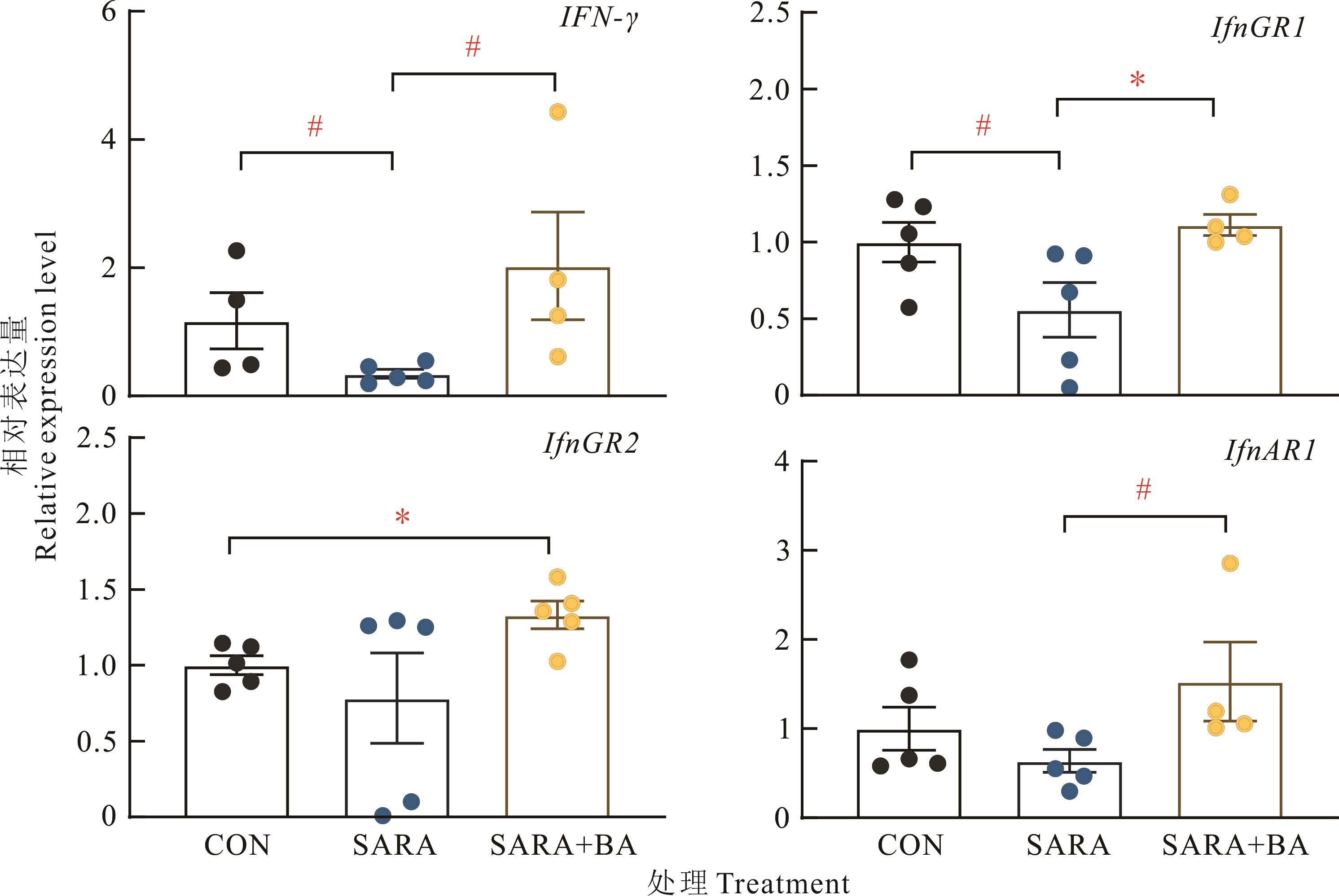
Fig.6 Effects of dietary supplementation with bile acids on the relative expression levels of interferon γ and its receptor mRNA in ileal mucosa of SARA goats (n=5)

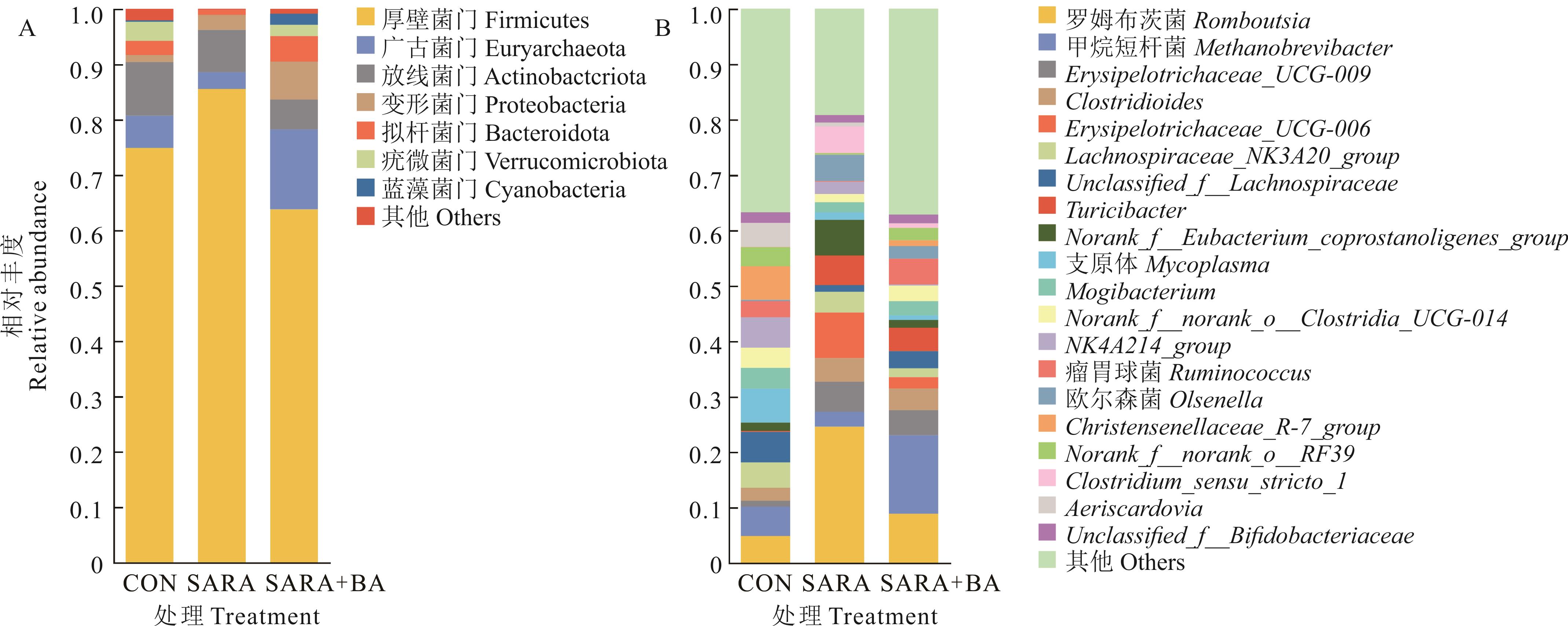
Fig.8 Effect of dietary supplementation with bile acids on the relative abundance at phylum level (A) and genus level (B) of ileal bacteria in SARA goats
| 1 | Ba L J D M, Ao R G L, Wang C J, et al. Pathogenesis and research progress of subacute rumen acidosis in ruminants. Feed Research, 2023, 46(21): 145-149. |
| 巴拉吉德玛, 敖日格乐, 王纯洁, 等. 反刍动物亚急性瘤胃酸中毒的发病机理及研究进展. 饲料研究, 2023, 46(21): 145-149. | |
| 2 | Dong G, Liu S, Wu Y, et al. Diet-induced bacterial immunogens in the gastrointestinal tract of dairy cows: Impacts on immunity and metabolism. Acta Veterinaria Scandinavica, 2011, 53(1): 48-55. |
| 3 | Acciarino A, Diwakarla S, Handreck J, et al. The role of the gastrointestinal barrier in obesity-associated systemic inflammation. Obesity Reviews, 2024, 25: e13673. |
| 4 | Elmhadi M E, Ali D K, Khogali M K, et al. Subacute ruminal acidosis in dairy herds: Microbiological and nutritional causes, consequences, and prevention strategies. Animal Nutrition, 2022, 10: 148-155. |
| 5 | Shi W R, Zhou F, Meng Q H, et al. Function and application in animal production of bile acids. Feed Research, 2023,46(15): 167-172. |
| 施文瑞, 周凡, 孟庆辉, 等. 胆汁酸的功能及其在动物生产中的应用. 饲料研究, 2023, 46(15): 167-172. | |
| 6 | He S Q, Li L X, Yao Y N, et al. Bile acid and its bidirectional interactions with gut microbiota: A review. Critical Reviews in Microbiology, 2023, doi: 10.1080/1040841X.2023.2262020. |
| 7 | Hegyi P, Maléth J, Walters J R, et al. Guts and gall: Bile acids in regulation of intestinal epithelial function in health and disease. Psychological Review, 2018, 98(4): 1983-2023. |
| 8 | Cai J, Rimal B, Jiang C, et al. Bile acid metabolism and signaling, the microbiota, and metabolic disease. Clinical Pharmacology & Therapeutics, 2022, doi: 10.1016/j.pharmthera.2022.108238. |
| 9 | Tao S Y. Effects of high-concentrate diet on hindgut epithelial barrier in lactating dairy goats and the mechanisms involved. Nanjing: Nanjing Agricultural University, 2018. |
| 陶诗煜. 高精料日粮对泌乳期奶山羊后段肠道上皮屏障的影响及其机制. 南京: 南京农业大学, 2018. | |
| 10 | Deng L Q. Make regular paraffin sections quickly. Journal of Hubei University for Nationalities (Medical Edition), 2001(4): 59. |
| 邓利群. 快速制作常规石蜡切片. 湖北民族学院学报(医学版), 2001(4): 59. | |
| 11 | Caporaso J G, Lauber C L, Walters W A, et al. Global patterns of 16S rRNA diversity at a depth of millions of sequences per sample. Proceedings of the National Academy of Sciences of the United States of America, 2011, 108(S1): 4516-4522. |
| 12 | Plaizier J C, Danesh M M, Derakhshani H, et al. Review: Enhancing gastrointestinal health in dairy cows. Animal, 2018, 12(S2): 399-418. |
| 13 | Camilleri M, Madsen K, Spiller R, et al. Intestinal barrier function in health and gastrointestinal disease. Neurogastroenterology and Motility, 2012, 24(6): 503-512. |
| 14 | Chelakkot C, Ghim J, Ryu S H. Mechanisms regulating intestinal barrier integrity and its pathological implications. Experimental and Molecular Medicine, 2018, 50(8): 1-9. |
| 15 | Tao S Y, Duan M L, Dong H B, et al. High concentrate diet induced mucosal injuries by enhancing epithelial apoptosis and inflammatory response in the hindgut of goats. PLoS One, 2014, 9(10): e111596. |
| 16 | Lai Z, Lin L, Zhang J, et al. Effects of high-grain diet feeding on mucosa-associated bacterial community and gene expression of tight junction proteins and inflammatory cytokines in the small intestine of dairy cattle. Journal of Dairy Science, 2022, 105(8): 6601-6615. |
| 17 | Huai Y Y, Liang Z S, Ji S L, et al. Effects of recombinant epidermal growth factor on the proliferation of the intestinal epithelial cells in early-weaned pigs. Animal Husbandry and Veterinary Medicine, 2016, 48(4): 70-75 . |
| 槐玉英, 梁梓森, 纪少丽, 等. 重组表皮生长因子对仔猪小肠上皮细胞增殖活性的影响. 畜牧与兽医, 2016, 48(4): 70-75. | |
| 18 | Guo Y S, Yan S M, Shi B L, et al. Effects of Lactobacillus fermentum supplementation on the small intestinal villus structure of broilers. Chinese Journal of Animal Nutrition, 2011, 23(7): 1194-1200. |
| 郭元晟, 闫素梅, 史彬林, 等. 发酵乳酸杆菌对肉鸡小肠绒毛形态的影响. 动物营养学报, 2011, 23(7): 1194-1200. | |
| 19 | Allaire J M, Crowley S M, Law H T, et al. The intestinal epithelium: Central coordinator of mucosal immunity. Trends in Immunology, 2018, 39(9): 677-696. |
| 20 | Kim J J, Khan W I. Goblet cells and mucins: Role in innate defense in enteric infections. Pathogens, 2013, 2(1): 55-70. |
| 21 | Okumura R, Takeda K. Roles of intestinal epithelial cells in the maintenance of gut homeostasis. Experimental and Molecular Medicine, 2017, 49(5): e338. |
| 22 | van der Flier L G, Clevers H. Stem cells, self-renewal, and differentiation in the intestinal epithelium. Annual Review of Physiology, 2009, 71: 241-260. |
| 23 | Johansson M E, Hansson G C. Immunological aspects of intestinal mucus and mucins. Nature Reviews Immunology, 2016, 16(10): 639-649. |
| 24 | Cui C, Li L, Wu L, et al. Paneth cells in farm animals: Current status and future direction. Journal of Animal Science and Biotechnology, 2023, 14(1): 118. |
| 25 | Hou Q, Huang J, Ayansola H, et al. Intestinal stem cells and immune cell relationships: Potential therapeutic targets for inflammatory bowel diseases. Frontiers in Immunology, 2021, doi: 10.3389/fimmu.2020.623691. |
| 26 | Günther C, Martini E, Wittkopf N, et al. Caspase-8 regulates tnf-α-induced epithelial necroptosis and terminal ileitis. Nature, 2011, 477(7364): 335-339. |
| 27 | Ergun E, Ergun L, Asti R N, et al. Light and electron microscopic morphology of paneth cells in the sheep small intestine. Revue De Medecine Veterinaire, 2003, 154(5): 351-355. |
| 28 | El Bougrini J, Pampin M, Chelbi-Alix M K. Arsenic enhances the apoptosis induced by interferon gamma: Key role of IRF-1. Cellular and Molecular Biology (Noisy-le-grand), 2006, 52(1): 9-15. |
| 29 | Gocher A M, Workman C J, Vignali D A A. Interferon-gamma: Teammate or opponent in the tumour microenvironment? Nature Reviews Immunology, 2022, 22(3): 158-172. |
| 30 | Richard N, Savoye G, Leboutte M, et al. Crohn’s disease: Why the ileum? World Journal of Gastroenterolog, 2023, 29(21): 3222-3240. |
| 31 | Thoetkiattikul H, Mhuantong W, Laothanachareon T, et al. Comparative analysis of microbial profiles in cow rumen fed with different dietary fiber by tagged 16S rRNA gene pyrosequencing. Current Microbiology, 2013, 67(2): 130-137. |
| 32 | Plaizier J C, Li S, Danscher A M, et al. Changes in microbiota in rumen digesta and feces due to a grain-based subacute ruminal acidosis (SARA) challenge. Microbial Ecology, 2017, 74(2): 485-495. |
| 33 | Zhong X X, Huang J, Liu Z Y, et al. Effects of mannan oligosaccharides and complex probiotics on growth performance and intestinal morphology,volatile fatty acid contents and microbial structure of weaned piglets. Chinese Journal of Animal Nutrition, 2020, 32(7): 3099-3108. |
| 钟晓霞, 黄健, 刘志云, 等. 甘露寡糖和复合益生菌对断奶仔猪生长性能及肠道形态结构、挥发性脂肪酸含量和菌群结构的影响. 动物营养学报, 2020, 32(7): 3099-3108. | |
| 34 | Crost E H, Coletto E, Bell A, et al. Ruminococcus gnavus: Friend or foe for human health. FEMS Microbiology Reviews, 2023, https://doi.org/10.1093/femsre/fuad014. |
| 35 | Zeng Y, Gao Y H, Peng Z L, et al. Effects of yeast culture supplementation in diets on rumen fermentation parameters and microflora of house-feeding yak. Chinese Journal of Animal Nutrition, 2020, 32(4): 1721-1733. |
| 曾钰, 高彦华, 彭忠利, 等. 饲粮中添加酵母培养物对舍饲牦牛瘤胃发酵参数及微生物区系的影响. 动物营养学报, 2020, 32(4): 1721-1733. | |
| 36 | Guo J, Mu R, Li S, et al. Characterization of the bacterial community of rumen in dairy cows with laminitis. Genes, 2021, doi: 10.3390/genes12121996. |
| 37 | Wang K, Ren A, Zheng M, et al. Diet with a high proportion of rice alters profiles and potential function of digesta-associated microbiota in the ileum of goats. Animals, 2020, doi: 10.3390/ani10081261. |
| 38 | Chiang J Y. Bile acid metabolism and signaling. Comprehensive Physiology, 2013, 3(3): 1191-1212. |
| 39 | Fogelson K A, Dorrestein P C, Zarrinpar A, et al. The gut microbial bile acid modulation and its relevance to digestive health and diseases. Gastroenterology, 2023, 164(7): 1069-1085. |
| [1] | Ming-ming GU, Xing-hui JIANG, Zhi-yi MA, Shui-ling QIU, Hao-yu LIU, Ming-rui ZHANG, Jia-ning LU, Yu-jun QIU, Ben-zhi WANG, Qian-fu GAN. Degradation characteristics of sweet potato and taro in the rumen of Mindong goats and changes in microbial community attached to the surface [J]. Acta Prataculturae Sinica, 2024, 33(9): 169-184. |
| [2] | Li-ping HONG, Xiao-dong LI, Er-ru YU, Cheng-jiang PEI, Yi-shun SHANG, Jin-hong LUO, Guang SUN, Yun-hao ZHOU, Shi-ge LI, Hang YANG, Feng-dan LIU. Effects of different perilla (Perilla frutescens) materials on serum antioxidant enzyme activity, rumen fermentation parameters and microflora of Guizhou black goats [J]. Acta Prataculturae Sinica, 2024, 33(9): 214-226. |
| [3] | Shang-lin YANG, Xuan WU, Qiao-hui LUO, Tai-hua HUANG, Zheng-fan ZHANG, Hai-tao SHI, Chun-hua GUO. A study of the protein requirements of 20-35 kg Chuanzhong black goats [J]. Acta Prataculturae Sinica, 2024, 33(7): 119-129. |
| [4] | Jie ZHAO, Heng-guang CHEN, Xiao-meng PEI, Hao YU, Yin-ying XU, Da-gan MAO. Effects of resveratrol supplementation in the perinatal diet on production performance, blood indexes, and transcript abundance of genes encoding inflammatory factors in goats [J]. Acta Prataculturae Sinica, 2024, 33(4): 210-220. |
| [5] | Tao ZHANG, Ying-yu MU, Wang-pan QI, Ji-you ZHANG, Sheng-yong MAO. Comparison of rumen epithelium morphology and function in dairy cows with differences in susceptibility for subacute ruminal acidosis [J]. Acta Prataculturae Sinica, 2023, 32(2): 131-139. |
| [6] | Jun-nian LI, Shao-hua KANG, Dong-mei YANG, Qian HE, Shuang LI, Shuang-lun TAO. Effects of substituting dietary alfalfa meal with kudzu vine (Pueraria lobata) meal on serum biochemical indexes, apparent nutrient digestibility and growth performance in Boer crossbred goats [J]. Acta Prataculturae Sinica, 2021, 30(8): 146-153. |
| [7] | Tao ZHANG, Ying-yu MU, Wang-pan QI, Chang-zheng GUO, Ji-you ZHANG, Sheng-yong MAO. Analysis of plasma and milk fatty acid and metabolite composition in lactating dairy cows with differing tolerance to subacute ruminal acidosis [J]. Acta Prataculturae Sinica, 2021, 30(7): 101-110. |
| [8] | Wang-bin SUN, Qi FU, Rui-lin XUE, Wei-ping WANG, Qian ZHANG, Ping FENG. Effects of different levels of jujube powder on slaughter characteristics and meat quality of Northern Shaanxi white cashmere goats [J]. Acta Prataculturae Sinica, 2021, 30(7): 111-121. |
| [9] | Li-qin HUANG, Song-qiao LI, Zhen-zhong YUAN, Jing TANG, Jing-cai YAN, Qi-yuan TANG. Effects of feeding co-fermented whole plant rice and spent mushroom (Pleurotus ostreatus) substrate on slaughter performance, meat quality and organ size indexes of Liuyang black goats [J]. Acta Prataculturae Sinica, 2021, 30(6): 133-140. |
| [10] | Jun-hong HUO, Kang ZHAN, Qiu-sheng HUANG, Xiao-jun ZHONG, Jin-shun ZHAN, Xue-bing YAN. Effects of different concentration∶roughage ratios on growth performance, serum biochemical indices and ruminal fermentation of Nubian goats [J]. Acta Prataculturae Sinica, 2021, 30(6): 151-161. |
| [11] | Wang-pan QI, Ying-yu MU, Tao ZHANG, Ji-you ZHANG, Sheng-yong MAO. Plasma biochemical indexes and metabolomics profile changes of dairy cows with subacute ruminal acidosis [J]. Acta Prataculturae Sinica, 2021, 30(6): 141-150. |
| [12] | Mang-li XIONG, Xu-jin WU, Xiao-fu ZHU, Wen-juan ZHANG. Effects of different apple pomace levels on lactation performance, nutrient apparent digestibility, serum biochemical indices and the rumen pH of Guanzhong dairy goats [J]. Acta Prataculturae Sinica, 2021, 30(3): 81-88. |
| [13] | Ji-qing WANG, Ji-yuan SHEN, Xiu LIU, Shao-bin LI, Yu-zhu LUO, Meng-li ZHAO, Zhi-yun HAO, Na KE, Yi-ze SONG, Li-rong QIAO. Comparative analysis of meat production traits, meat quality, and muscle nutrient and fatty acid contents between Ziwuling black goats and Liaoning cashmere goats [J]. Acta Prataculturae Sinica, 2021, 30(2): 166-177. |
| [14] | Chen WU, Zhi-hao YAO, Wen-qing MEI, Yu-yan FENG, Qu CHEN, Ying-dong NI. Effects of vitamin B complex on intestinal microflora composition and gut epithelial structure in growing goats [J]. Acta Prataculturae Sinica, 2021, 30(11): 170-180. |
| [15] | ZHAN Jin-shun, HUO Jun-hong, HU Yao, ZHONG Xiao-jun, WU Yan-ping. Effects of total mixed rations with different concentrate∶roughage ratios on meat quality, serum indexes and organ development in Nubian goats [J]. Acta Prataculturae Sinica, 2020, 29(10): 139-148. |
| Viewed | ||||||
|
Full text |
|
|||||
|
Abstract |
|
|||||