
ISSN 1004-5759 CN 62-1105/S
Acta Prataculturae Sinica ›› 2021, Vol. 30 ›› Issue (10): 92-104.DOI: 10.11686/cyxb2020360
Previous Articles Next Articles
Kai YANG( ), Juan SHI(), Yu-tao YUAN, Li-ting WANG
Received:2020-07-28
Revised:2020-09-27
Online:2021-09-16
Published:2021-09-16
Contact:
Juan SHI
Kai YANG, Juan SHI, Yu-tao YUAN, Li-ting WANG. Pathogen identification and cell physiological changes of Trifolium repens leaves infected with powdery mildew[J]. Acta Prataculturae Sinica, 2021, 30(10): 92-104.

Fig. 1 Symptoms of leaf damage of T. repens by E. pisi
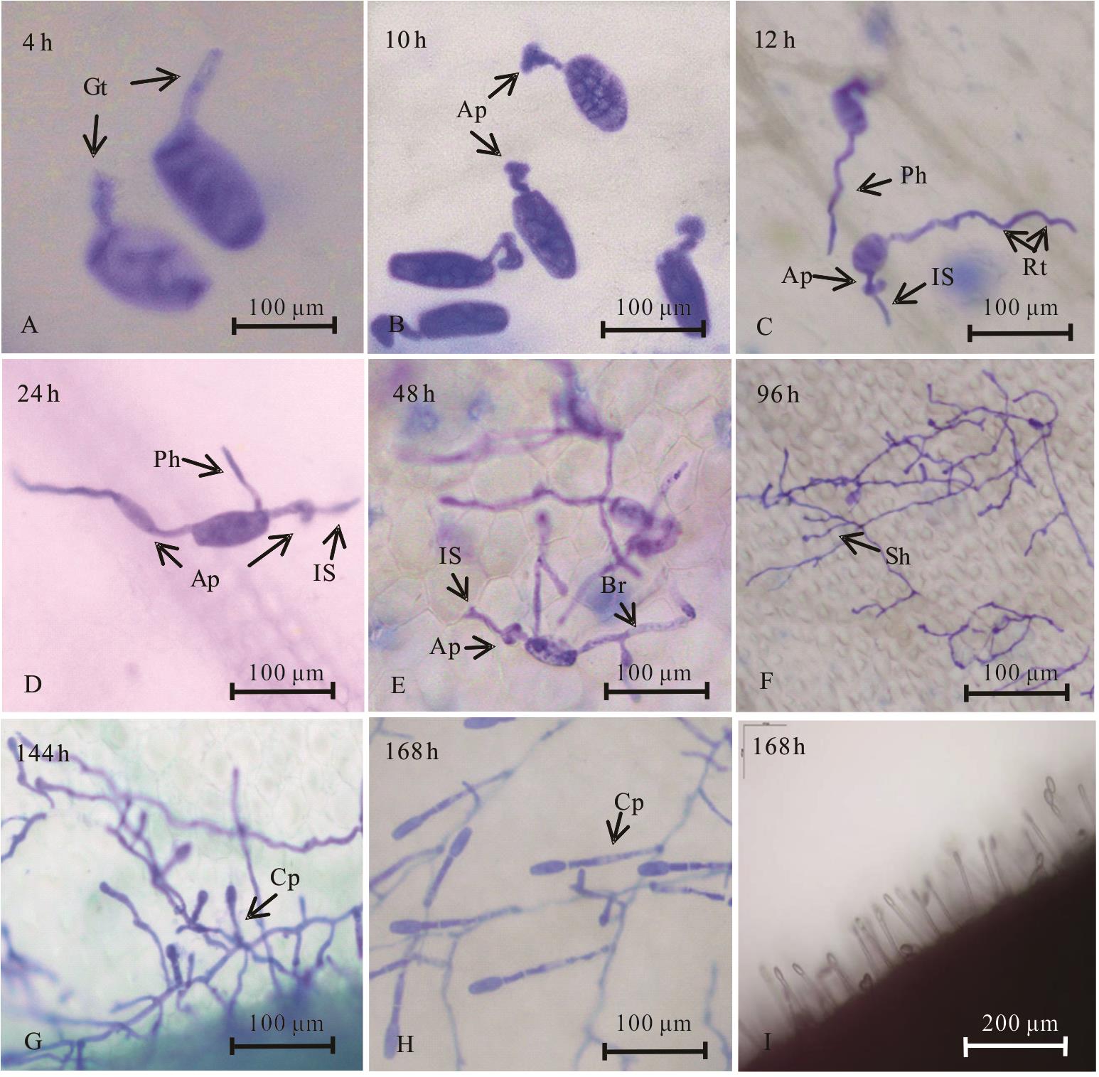
Fig. 2 Development of E. pisi in T. repens

Fig. 3 A semi-thin section of normal T. repens host tissue and powdery mildew invaded the host tissue

Fig. 4 Ultrastructural characteristics of pathogen invading host cells
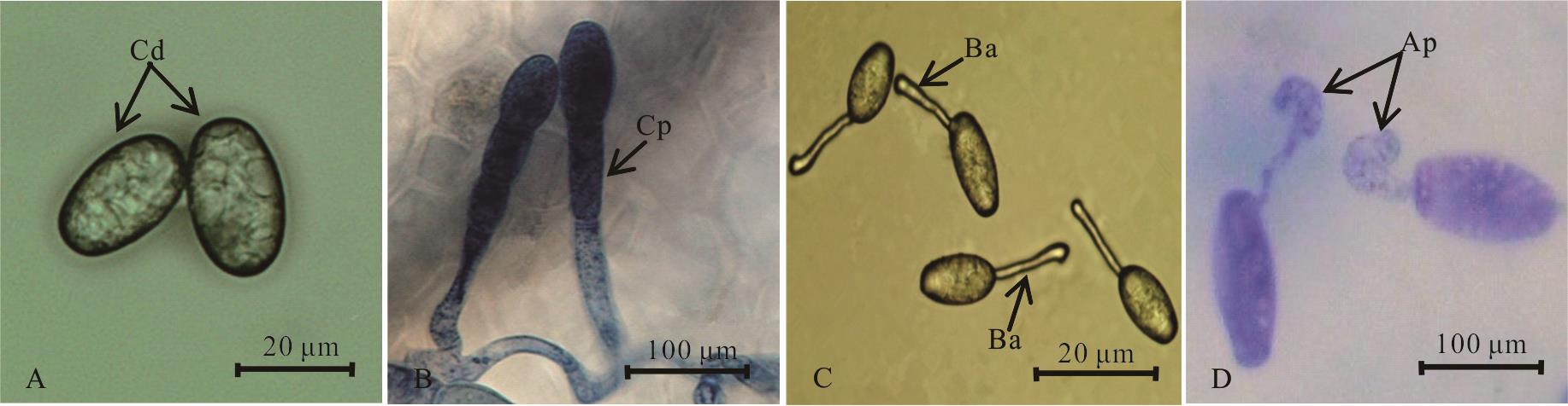
Fig.5 Morphological characteristics of fungus causing powdery mildews on T. repens
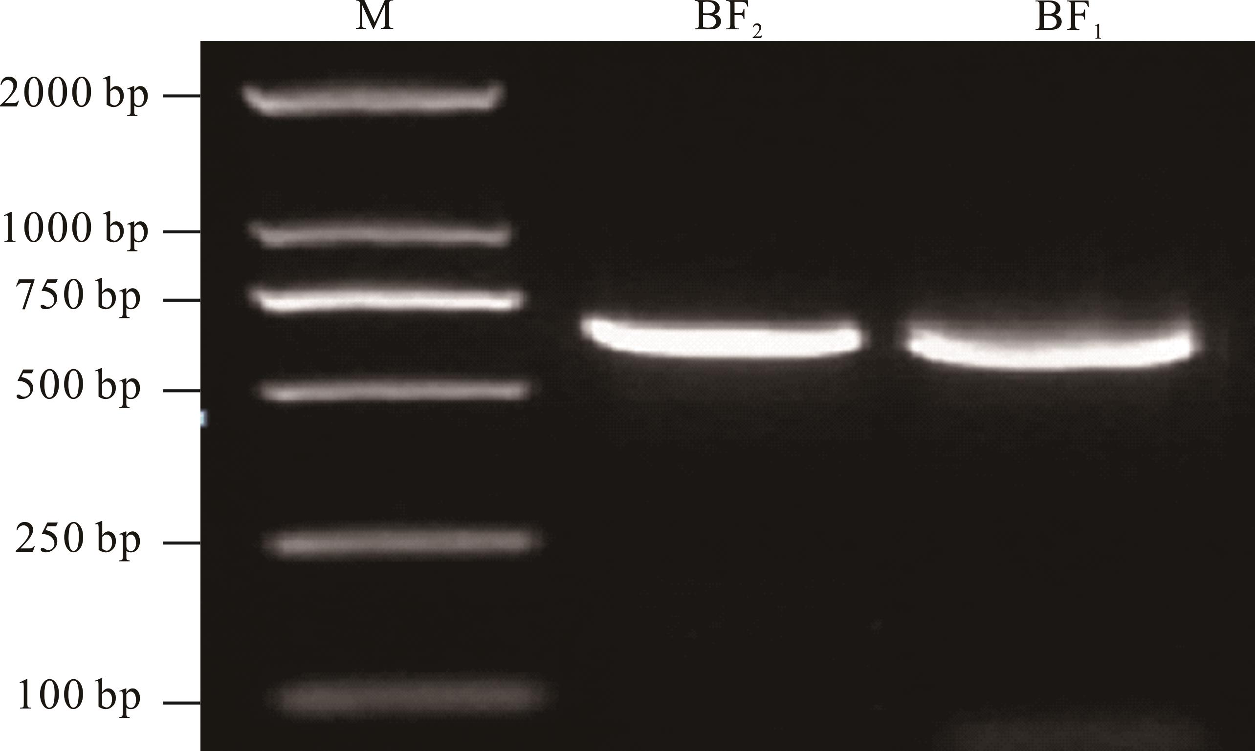
Fig.6 PCR amplification of ribosomal ITS sequence segments of powdery mildew

Fig.7 Phylogenetic tree constructed by ITS sequence
温度 Temperature (℃) | 孢子萌发率 Spore germination rate (%) | ||||
|---|---|---|---|---|---|
| 4 h | 6 h | 8 h | 12 h | 24 h | |
| 5 | 0.53C | 2.17C | 2.97B | 5.45B | 7.55B |
| 10 | 0.98C | 4.77C | 8.27B | 16.00B | 17.43B |
| 15 | 7.01B | 19.00B | 27.40A | 33.15A | 40.47A |
| 20 | 9.42AB | 18.55B | 21.50A | 32.29A | 35.85A |
| 25 | 11.57A | 28.21A | 31.25A | 37.76A | 47.53A |
| 28 | 2.54C | 8.80C | 9.63B | 14.83B | 18.65B |
| 30 | 0.97C | 4.62C | 6.78B | 7.19B | 9.15B |
Table 1 Effects of different temperatures on the conidia germination of powdery mildew in T. repens
温度 Temperature (℃) | 孢子萌发率 Spore germination rate (%) | ||||
|---|---|---|---|---|---|
| 4 h | 6 h | 8 h | 12 h | 24 h | |
| 5 | 0.53C | 2.17C | 2.97B | 5.45B | 7.55B |
| 10 | 0.98C | 4.77C | 8.27B | 16.00B | 17.43B |
| 15 | 7.01B | 19.00B | 27.40A | 33.15A | 40.47A |
| 20 | 9.42AB | 18.55B | 21.50A | 32.29A | 35.85A |
| 25 | 11.57A | 28.21A | 31.25A | 37.76A | 47.53A |
| 28 | 2.54C | 8.80C | 9.63B | 14.83B | 18.65B |
| 30 | 0.97C | 4.62C | 6.78B | 7.19B | 9.15B |
光照时间 Illumination treatment | 孢子萌发率Spore germination rate | ||||
|---|---|---|---|---|---|
| 4 h | 6 h | 8 h | 12 h | 24 h | |
| 全光照 Full illumination | 20.06A | 36.88A | 40.75A | 43.40A | 45.82A |
| 光暗交替 Half illumination | 13.90AB | 27.54A | 30.12A | 36.66A | 39.96A |
| 全黑暗 Total darkness | 8.33B | 20.98A | 27.03A | 30.17A | 33.87A |
Table 2 Effect of different lights on the conidia germination of powdery mildew in T. repens (%)
光照时间 Illumination treatment | 孢子萌发率Spore germination rate | ||||
|---|---|---|---|---|---|
| 4 h | 6 h | 8 h | 12 h | 24 h | |
| 全光照 Full illumination | 20.06A | 36.88A | 40.75A | 43.40A | 45.82A |
| 光暗交替 Half illumination | 13.90AB | 27.54A | 30.12A | 36.66A | 39.96A |
| 全黑暗 Total darkness | 8.33B | 20.98A | 27.03A | 30.17A | 33.87A |
| pH | 孢子萌发率Spore germination rate | ||||
|---|---|---|---|---|---|
| 4 h | 6 h | 8 h | 12 h | 24 h | |
| 4.0 | 2.75CD | 10.87BC | 12.54C | 15.20B | 15.78B |
| 5.0 | 2.94CD | 11.12BC | 14.25B | 19.66B | 25.82B |
| 6.0 | 11.08B | 17.73AB | 23.20B | 32.43A | 37.51A |
| 7.0 | 16.86A | 24.77A | 35.97A | 40.99A | 45.50A |
| 8.0 | 8.01BC | 15.02B | 23.11B | 31.56A | 37.12A |
| 9.0 | 2.13D | 4.95C | 7.50C | 12.75B | 18.72B |
Table 3 Effects of different pH on the conidia germination of powdery mildew in T. repens (%)
| pH | 孢子萌发率Spore germination rate | ||||
|---|---|---|---|---|---|
| 4 h | 6 h | 8 h | 12 h | 24 h | |
| 4.0 | 2.75CD | 10.87BC | 12.54C | 15.20B | 15.78B |
| 5.0 | 2.94CD | 11.12BC | 14.25B | 19.66B | 25.82B |
| 6.0 | 11.08B | 17.73AB | 23.20B | 32.43A | 37.51A |
| 7.0 | 16.86A | 24.77A | 35.97A | 40.99A | 45.50A |
| 8.0 | 8.01BC | 15.02B | 23.11B | 31.56A | 37.12A |
| 9.0 | 2.13D | 4.95C | 7.50C | 12.75B | 18.72B |
| 1 | Wang Z Y, Shu J H, Chen Y, et al.Analysis of Trifolium repens DNA methylation of Guizhou different regions by MSAP. Genomics and Applied Biology, 2020, 39(4): 1741-1750. |
| 王子苑, 舒健虹, 陈莹, 等. 贵州不同地区白三叶基因组甲基化MSAP分析. 基因组学与应用生物学, 2020, 39(4): 1741-1750. | |
| 2 | Huang Y M, Zhang K, Sun L X, et al.Effects of Trifolium repens invasion on soil animals in an urban turf ecosystem. Acta Ecologica Sinica, 2018, 38(23): 8489-8499. |
| 黄玉梅, 张凯, 孙凌霞, 等. 白三叶(Trifolium repens)入侵对城市草坪生态系统土壤动物的影响. 生态学报, 2018, 38(23): 8489-8499. | |
| 3 | Liang Z, Jiang S J, Wei L, et al. Progress in research on Trifolium genetic engineering. Acta Prataculturae Sinica, 2009, 18(2): 205-211. |
| 梁哲, 姜三杰, 未丽, 等. 三叶草基因工程研究进展. 草业学报, 2009, 18(2): 205-211. | |
| 4 | Chen B S. Forage crop cultivation. Beijing: China Agriculture Press, 2001: 224-233. |
| 陈宝书. 牧草饲料作物栽培学. 北京: 中国农业出版社, 2001: 224-233. | |
| 5 | Li Y X, Zhang T B, Du H L, et al. The allelopathy of five pine needle extracts on Trifolium repens L. Journal of Nuclear Agricultural Sciences, 2020, 34(7): 1606-1612. |
| 李雅馨, 张天宝, 杜慧玲, 等. 5种松针浸提液对白三叶草的化感作用. 核农学报, 2020, 34(7): 1606-1612. | |
| 6 | Jin Z M, Sha W. Study on drought resistance of Trifolium repens Linn seedlings. Northern Horticulture, 2010(18): 50-52. |
| 金忠民, 沙伟. 白三叶抗旱生理的研究. 北方园艺, 2010(18): 50-52. | |
| 7 | Li Z, Peng Y, Yin S X, et al. Effects of exogenous mannose application on drought tolerance, sugars, and sugar alcohol accumulation in white clover. Acta Prataculturae Sinica, 2019, 28(12): 85-93. |
| 李州, 彭燕, 尹淑霞, 等. 甘露糖对白三叶抗旱性、糖及糖醇类代谢物积累的影响. 草业学报, 2019, 28(12): 85-93. | |
| 8 | Qu L L, Wang J J. Research progress on stress resistance of Trifolium repens. Chinese Journal of Grassland, 2020, 42(2): 155-161. |
| 屈璐璐, 王俊杰. 白三叶抗逆性研究进展. 中国草地学报, 2020, 42(2): 155-161. | |
| 9 | Lan H Y. Common diseases and insect pests of herbage and their control methods. Contemporary Animal Husbandry, 2018(26): 17-18. |
| 兰惠英. 牧草常见病虫害及防治方法. 当代畜牧, 2018(26): 17-18. | |
| 10 | Liu X L, Song C, Du W H. Study on mechanism of resistance to powdery mildew in Minshan red clover. Grassland and Turf, 2011, 31(3): 73-76. |
| 刘晓玲, 宋超, 杜文华. 红三叶对白粉病抗性机理研究. 草原与草坪, 2011, 31(3): 73-76. | |
| 11 | Yang F. Identification and prevention of powdery mildew in garden lawn. Xinjiang Agricultural Science and Technology, 2008(3): 62. |
| 杨芬. 园林草坪白粉病的识别与防治. 新疆农业科技, 2008(3): 62. | |
| 12 | Sang W J, Liang J, Li Y S. Investigation on powdery mildew inlidence of Trifolium pratense in Guiyang. Journal of Southwest University (Natural Science Edition), 1990(4): 353-355. |
| 桑维均, 梁建, 李永顺. 贵阳地区红三叶草白粉病病情消长和病菌生物学特性. 西南农业大学学报(自然科学版), 1990(4): 353-355. | |
| 13 | Yuan Y T, Shi J, Ma X, et al. Identification and biological characteristics of alfalfa powdery mildew fungi. Microbiology China, 2020, 47(11): 3539-3550. |
| 袁玉涛, 史娟, 马新, 等. 紫花苜蓿白粉病病原菌鉴定及其生物学特性. 微生物学通报, 2020, 47(11): 3539-3550. | |
| 14 | Li S, Zhu L L, Li J F, et al. Identification of the pathogen causing tomato powdery mildew in Heilongjiang Province. Plant Protection, 2014, 40(4): 112-114, 129. |
| 李帅, 朱路路, 李景富, 等. 黑龙江省番茄白粉病病原鉴定. 植物保护, 2014, 40(4): 112-114, 129. | |
| 15 | Zheng L, Wu X Q.Advances on infection structures of plant pathogenic fungi. Journal of Nanjing Forestry University (Natural Sciences Edition) , 2007(1): 90-94. |
| 郑玲, 吴小芹. 植物病原真菌侵染结构研究进展. 南京林业大学学报(自然科学版), 2007(1): 90-94. | |
| 16 | Riopel J L. Haustorial initiation and differentiation. New York: Parasitic Plants, Chapman and HallPress, 1995. |
| 17 | Ren B, Gao X N, Han Q M, et al. Etiology and infection process of Glomerella cingulata causing Glomerella leaf spot of apple. Acta Phytophylacica Sinica, 2014, 41(5): 608-614. |
| 任斌, 高小宁, 韩青梅, 等. 苹果炭疽叶枯病病原(Glomerella cingulata)及其侵染过程. 植物保护学报, 2014, 41(5): 608-614. | |
| 18 | Yang R L, Liu J Y, Lv X, et al.Micro-and ultrastructural changes of wheat leaf cells induced by powdery mildew infection. Acta Botanica Boreali-Occidentalia Sinica, 2001(2): 293-296, 400-401. |
| 杨若林, 刘建云, 吕欣, 等. 白粉菌侵染对小麦叶片显微及超微结构的影响. 西北植物学报, 2001(2): 293-296, 400-401. | |
| 19 | Zhang Z L, Qu W J. Experimental instruction in plant physiology (3rd Edition). Beijing: Higher Education Press, 2003: 206. |
| 张志良, 瞿伟菁. 植物生理学实验指导(第3版). 北京: 高等教育出版社, 2003: 206. | |
| 20 | Kang Z S. Ultrastructure of plant pathogenic fungi. Beijing: China Science and Technology Press, 1996: 1-74. |
| 康振生. 植物病原真菌超微结构. 北京: 中国科学技术出版社, 1996: 1-74. | |
| 21 | Li W X, Xiao R G, Lv M M, et al. Establishment and application of real-time PCR for quantitatively detecting Plasmopara viticola in Vitis vinifera. Scientia Agricultura Sinica, 2019, 52(9): 1529-1540. |
| 李文学, 肖瑞刚, 吕苗苗, 等. 葡萄霜霉病菌实时荧光定量PCR检测体系的建立和应用. 中国农业科学, 2019, 52(9): 1529-1540. | |
| 22 | Fang Z D. Research methods of plant diseases. Beijing: China Agriculturae Press, 1998: 140-145. |
| 方中达. 植病研究方法. 北京: 中国农业出版社, 1998: 140-145. | |
| 23 | Wei J C. Fungus identification manual. Shanghai: Shanghai Scientific & Technical Publishers, 1979: 1-9. |
| 魏景超. 真菌鉴定手册. 上海: 上海科学技术出版社, 1979: 1-9. | |
| 24 | Zheng R Y, Yu Y N. Flora of fungi in China, Volume I, powdery mildew. Beijing: Science Press, 1987: 5-13. |
| 郑儒永, 余永年. 中国真菌志, 第一卷, 白粉菌目. 北京: 科学出版社, 1987: 5-13. | |
| 25 | Bushnell W R. Aggregation of host cytoplasm and the formation of papillae and haustoria in powdery mildew of barley. Phytopathology, 1975, 65: 310-318. |
| 26 | Wan S L, Liang P, Liu W B, et al.Cytological analysis of compatible interactions between rubber tree and Oidium heveae. Plant Protection, 2014, 40(3): 26-36. |
| 万三连, 梁鹏, 刘文波, 等. 橡胶树与白粉病菌(Oidium heveae)亲和互作组织细胞学研究. 植物保护, 2014, 40(3): 26-36. | |
| 27 | Zhang Y M, Ma H L, Tang Y Z. Structural changes in leaves in Medicago sativa infected with Erysiphe pisi. Acta Prataculturae Sinica, 2017, 26(2): 88-94. |
| 张咏梅, 马晖玲, 唐云智. 紫花苜蓿叶片受白粉病菌侵染后结构的变化. 草业学报, 2017, 26(2): 88-94. | |
| 28 | Lu Q, Yang L, Wang H W, et al.Responses of photosynthetic characteristics and chloroplast ultrastructure to salt stress in seedlings of Cornus hongkongensis subsp. elegans. Journal of Nanjing Forestry University (Natural Sciences Edition), 2020, 44(4): 29-36. |
| 鲁强, 杨玲, 王昊伟, 等. 秀丽四照花光合特性和叶绿体超微结构的盐胁迫响应. 南京林业大学学报(自然科学版), 2020, 44(4): 29-36. | |
| 29 | Li X F, Ni Z M, Wu Y Y, et al.Effects of salt stress on photosynthetic characteristics and leaf cell structure of ‘Yinhong’ grape seedlings. Acta Ecologica Sinica, 2015, 35(13): 4436-4444. |
| 李学孚, 倪智敏, 吴月燕, 等. 盐胁迫对‘鄞红’葡萄光合特性及叶片细胞结构的影响. 生态学报, 2015, 35(13): 4436-4444. | |
| 30 | Kirchhoff H . Chloroplast ultrastructure in plants. New Phytologist, 2019, 223(2): 565-574. |
| 31 | Van Wijk K J, Kessler F. Plastoglobuli: Plastid microcompartments with integrated functions in metabolism, plastid developmental transitions, and environmental adaptation. Annual Review of Plant Biology, 2017, 68(1): 253-289. |
| 32 | Liu M, Fang Y L. Effects of heat stress on physiological indexes and ultrastructure of grapevines. Scientia Agricultura Sinica, 2020, 53(7): 1444-1458. |
| 刘敏, 房玉林. 高温胁迫对葡萄幼树生理指标和超显微结构的影响. 中国农业科学, 2020, 53(7): 1444-1458. | |
| 33 | Xu W, Wang Y, Yuan Q H, et al. Effect of NaCl on ultrastructure in leaves of white clovers (Trifolium repens L.). Chinese Journal of Grassland, 2013, 35(6): 104-108. |
| 徐威, 王瑜, 袁庆华, 等. NaCl胁迫对白三叶叶片超微结构的影响. 中国草地学报, 2013, 35(6): 104-108. | |
| 34 | Shi J, Wang H R, Zhong S L.Ultrastructural characteristics of compatible Pseudopeziza medicaginis interaction with alfalfa leaf. Acta Prataculturae Sinica, 2012, 21(5): 122-127. |
| 史娟, 王华荣, 钟少林. 苜蓿假盘菌与苜蓿叶片亲和性互作的超微结构特征. 草业学报, 2012, 21(5): 122-127. | |
| 35 | Chen S H, Lu Z Q. Occurrence and control measures of powdery mildew of red clover in artificial pasture. Journal of Grassland and Forage Science, 1997(2): 47-48. |
| 陈曙晖, 陆忠权. 人工草场红三叶草白粉病的发生与防治措施. 四川草原, 1997(2): 47-48. | |
| 36 | Yan Y, Li W P, Gao W J, et al. Application of rDNA ITS sequence analysis in fungus identification. Chinese Journal of Health Laboratory Technology, 2008(10): 1958-1961. |
| 燕勇, 李卫平, 高雯洁, 等. rDNA-ITS序列分析在真菌鉴定中的应用. 中国卫生检验杂志, 2008(10): 1958-1961. | |
| 37 | Wang L J, Guo N, Wang W, et al. The soil water content monitoring based on the temperature difference in the Northwest China. Research of Soil and Water Conservation, 2018, 25(5): 330-336. |
| 王丽娟, 郭铌, 王玮, 等. 基于温度差监测西北地区的土壤相对湿度. 水土保持研究, 2018, 25(5): 330-336. |
| [1] | WANG Qiong, DUAN Ting-yu, NAN Zhi-biao. Isolation and identification of an anthracnose pathogen on Vicia sativa [J]. Acta Prataculturae Sinica, 2020, 29(6): 127-136. |
| [2] | WANG Chun-ming, YUAN Wei-wei, ZHANG Xiao-jie, ZHOU Tian-wang, GUO Cheng, JIN She-lin. Isolation, identification and biological characteristics of Alternaria brassicicola leaf spot on Orychophragums violaceus [J]. Acta Prataculturae Sinica, 2020, 29(5): 88-97. |
| [3] | CHEN Bin, LI Hong-yao, LIU Xiao-wei, XIA Bin, SUN Shao-wen, SUN Ying, HE Miao. Effects of different light intensities on morphogenesis and ultrastructure of Gibasis pellucida leaf [J]. Acta Prataculturae Sinica, 2019, 28(7): 175-185. |
| [4] | SUN Hai-rong, CHE Zhao-bi, CHEN Yi-shi, LU Wei-hua, WANG Shu-lin, LI Na-na, XIN Huai-lu. Ecological adaptability of biological traits and population distribution patterns for the ephemeral plant Leontice incerta in desert habitats [J]. Acta Prataculturae Sinica, 2019, 28(7): 198-207. |
| [5] | LI Jian-hong, LI Xue-ping, LI Chang-ning, HAN Bing, XU Wan-li, YAO Tuo. Characterization of a plant-growth-promoting rhizosphere bacterium, Gnyt1, and determination of its taxonomic status [J]. Acta Prataculturae Sinica, 2019, 28(5): 55-67. |
| [6] | DING Ai-qiang, XÜ Xian-ying, ZHANG Wen, LIU Jiang, FU Li, FU Gui-quan. Soil physicochemical and biological characteristics of Tamarix ramosissima Nebkhas in different degradation degree [J]. Acta Prataculturae Sinica, 2019, 28(2): 1-11. |
| [7] | FENG Peng, SUN Li, SHEN Xiao-hui, LI Ru-lai, LI Zeng-jie, LI Zhi-min, ZHENG Hai-yan, JIANG Cheng, YNAG He, LIU Jun-gang, GUO Wei, ZHANG Ying-jun. Effect of different mutagenesis methods on microstructure and ultrastructure of alfalfa [J]. Acta Prataculturae Sinica, 2018, 27(6): 72-80. |
| [8] | PU Xiao-jian, TIAN Jiu-sheng, TIAN Xin-hui, DU Wen-hua. Construction of the AFLP linkage map and QTL analysis of powdery mildew resistance in red clover [J]. Acta Prataculturae Sinica, 2018, 27(4): 79-88. |
| [9] | YANG Cheng-De, BIAN Jing, CHEN Tai-Xiang, CHEN Xiu-Rong, WANG Han-Qi, YANG Xiao-Li, WANG Yang. Biological characteristics of the Angelica sinensis anthracnose causal agent, Colletotrichum dematium [J]. Acta Prataculturae Sinica, 2017, 26(6): 139-144. |
| [10] | HE Chun-Gui, HE Zhen-Fu, WANG Fei. Efficient double cropping pattern of photoperiod-sensitive sorghum-sudangrass hybrids in summer after winter wheat [J]. Acta Prataculturae Sinica, 2017, 26(5): 70-80. |
| [11] | LI Jian, LI Mei, GAO Xing-Xiang, FANG Feng, DONG Lian-Hong. Biological characteristics of Lubao No.1 biological control agent (Colletotrichum gloeosporioides) and construction of a T-DNA insertional mutant library [J]. Acta Prataculturae Sinica, 2017, 26(1): 142-148. |
| [12] | LI Jian, LI Mei, GAO Xing-Xiang, FANG Feng, DONG Lian-Hong. Isolation and biological characteristics of the biological control fungi BC-1 for Echinochloa crusgalli [J]. Acta Prataculturae Sinica, 2016, 25(8): 164-171. |
| [13] | ZHAO Fu-Qiang, ZHANG Hai-Qin, SUN Zong-Hua, JIAO Zhen-Fei, LIU Xiao-Yan, CHEN Wei-Huan, CHEN Guo-Yue, ZHOU Yong-Hong. Resistance of Roegneria kamoji (Poaceae: Triticeae) populations to stripe rust and powdery mildew [J]. Acta Prataculturae Sinica, 2016, 25(4): 149-158. |
| [14] | JIA Hui, CHEN Xiu-Rong, LU Guang-Xin, KONG Ya-Li, YANG Cheng-De. Isolation of cellulose-degrading bacteria and determination of their degradation activity [J]. Acta Prataculturae Sinica, 2016, 25(3): 60-66. |
| [15] | FANG Qiang-En, ZHANG Bo, LI Yu-Bo, SHI Shang-Li. Adaptive changes in the young leaf cell ultrastructure of crown buds in Medicago sativa (Leguminosae) during overwintering [J]. Acta Prataculturae Sinica, 2016, 25(3): 96-107. |
| Viewed | ||||||
|
Full text |
|
|||||
|
Abstract |
|
|||||